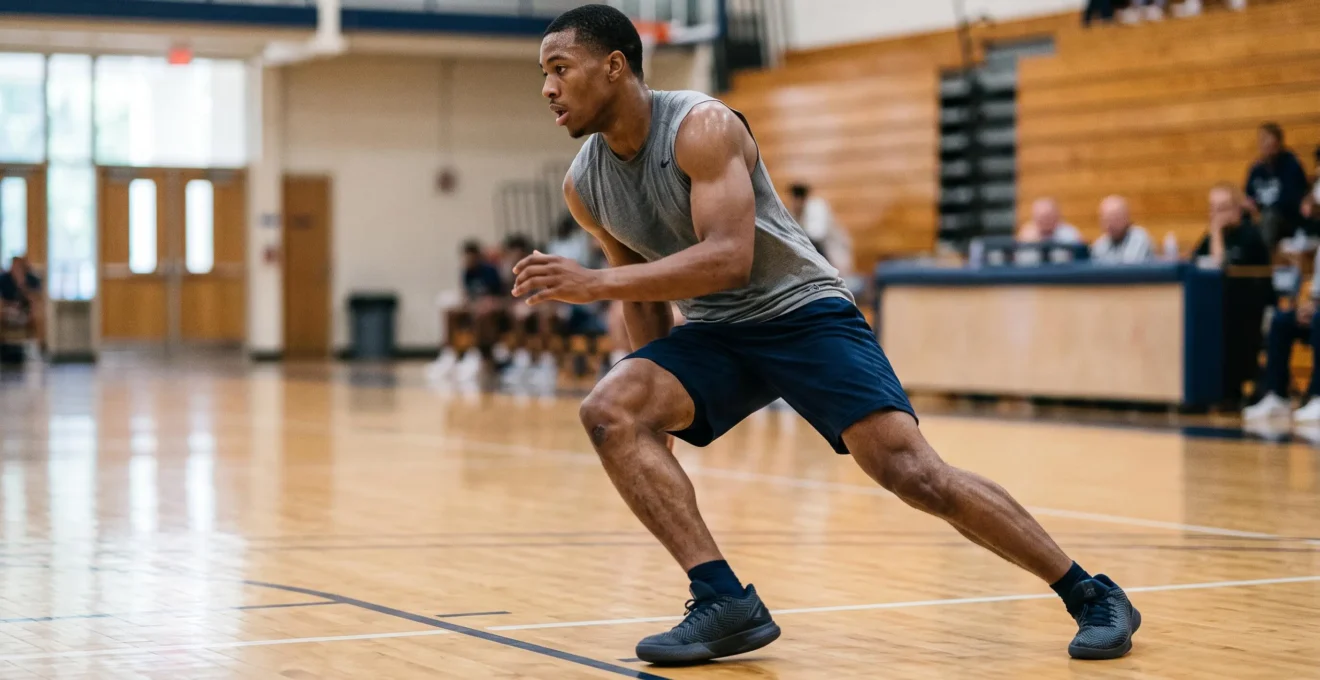
Three of Marcus’s training partners ended their season early due to knee injuries within two years. Two suffered ACL tears requiring surgery, months of rehabilitation, and uncertain return-to-sport timelines. As a semi-professional trail runner covering 80-100 kilometers weekly at altitude in Denver, Marcus now notices a persistent anterior knee discomfort after long runs—mild enough to disappear with rest, concerning enough to trigger questions about his training future and his eight-month timeline to qualify for the Ultra-Trail du Mont-Blanc.
This scenario reflects a broader challenge facing competitive athletes: distinguishing normal training adaptation from early warning signals while navigating contradictory prevention advice. Research demonstrates that structured neuromuscular training protocols can reduce knee injury risk by 30 to 50 percent, yet confusion persists about which interventions deliver measurable protection versus those offering marginal benefit. Effective prevention requires integrating three often-separated domains—biomechanical control, training architecture, and tissue recovery—rather than implementing isolated exercise lists without understanding individual risk profiles.
In this article
- The Injury Landscape: Why Knee Injuries Dominate Athletic Medicine
- Which biomechanical patterns elevate injury risk?
- Evidence-Based Neuromuscular Training Protocols
- How does movement quality impact long-term knee resilience?
- Recovery Architecture: Sleep and Tissue Adaptation
- When should athletes seek specialist evaluation?
- Building Your Integrated Prevention Framework
The Injury Landscape: Why Knee Injuries Dominate Athletic Medicine
0.23
per 1000 athlete-exposures
Highest average annual ACL injury rate among female collegiate athletes, recorded in lacrosse (NCAA Injury Surveillance System, 2004-2013)
Knee injuries represent the most frequent cause of prolonged sport interruption across disciplines involving rapid direction changes, deceleration forces, and repetitive landing cycles. NCAA surveillance data spanning 2004-2013 reveals striking variation in anterior cruciate ligament (ACL) tear incidence based on sport-specific biomechanical demands. Among male collegiate athletes, football recorded the highest rate at 0.17 injuries per 1000 athlete-exposures, while female lacrosse athletes experienced rates reaching 0.23 per 1000 exposures.
The data reveals a consistent pattern: women sustain ACL injuries at higher rates than men in comparable sports, including soccer, basketball, and lacrosse. This disparity reflects multiple contributing factors—anatomical differences in pelvic width and femoral angle, neuromuscular activation patterns during cutting and landing tasks, and hormonal influences on ligament laxity. Understanding these baseline risk profiles allows athletes to assess whether their chosen discipline places them in elevated-risk populations requiring proactive intervention.
Trail running and long-distance events present distinct injury mechanisms compared to pivot-dominant sports. While acute ACL tears remain less common in linear running disciplines, cumulative loading patterns generate anterior knee pain syndromes, patellar tendinopathy, and cartilage stress. Descents on technical terrain amplify eccentric loading demands, fatiguing the quadriceps muscles that normally protect the patellofemoral joint from excessive compression forces. For athletes like Marcus accumulating 100,000 ground contacts weekly, technical deficiencies multiplied across this volume create measurable tissue adaptation demands.
Beyond incidence rates, the long-term consequences of knee injuries justify prevention investment. Athletes who suffer ACL rupture face surgery, nine to twelve months of rehabilitation, elevated risk of contralateral injury, and accelerated development of post-traumatic osteoarthritis within 10-20 years. Return-to-sport rates following ACL reconstruction vary significantly, with substantial percentages of athletes never regaining their pre-injury competition level. These realities transform prevention from optional training supplement to career-preservation priority.
Navigating the complexities of high-level sports injuries often requires the guidance of recognized experts in orthopedic surgery. The work and surgical expertise of Prof. Etienne Cavaignac provide athletes with specialized insights into both preventive care and reconstructive procedures, ensuring that recovery protocols align with the latest biological and biomechanical standards. This clinical perspective is essential for those balancing intense performance goals with the necessity of maintaining joint integrity over a professional career.
Which biomechanical patterns elevate injury risk?
Biomechanical screening identifies movement deficits that predict injury before symptoms emerge. The most extensively documented pattern—dynamic knee valgus—describes medial collapse of the knee during weight-bearing activities. When viewed from the front during a landing or cutting maneuver, the knee moves inward relative to the foot and hip, creating a biomechanically disadvantaged position that concentrates stress on the ACL and medial knee structures.
Prospective research tracking athletes before injury occurrence demonstrates that excessive valgus motion during screening tests correlates with significantly higher ACL tear rates in subsequent seasons. This relationship exists because the inward knee position simultaneously increases ACL tension while reducing the joint’s capacity to dissipate ground reaction forces through muscle activation. Athletes exhibiting pronounced valgus during single-leg landings experience multifold increased injury risk compared to those maintaining neutral frontal plane alignment.
Landing mechanics represent the second critical assessment domain. Athletes who land from jumps with minimal knee flexion—maintaining relatively straight legs upon ground contact—generate peak force spikes that ligaments and cartilage must absorb without adequate muscular buffering. This rigid landing pattern, combined with asymmetrical left-right loading or excessively short ground contact times, signals deficient neuromuscular control strategies. Sports requiring repetitive jumping (basketball, volleyball) or aggressive downhill running (trail, ski) magnify the consequences of these technical deficiencies across thousands of loading cycles.
The third pattern involves proximal control deficits—specifically, inadequate hip abductor and external rotator strength allowing the femur to rotate inward and adduct during dynamic tasks. This hip-driven compensation cascades distally, forcing the knee into vulnerable positions. The confusion Marcus experiences regarding contradictory strengthening advice (quadriceps versus hamstrings versus hip musculature) reflects incomplete understanding of this kinetic chain relationship. Isolated quadriceps strengthening without addressing hip control and hamstring coordination fails to modify the movement patterns that generate injury risk.
Self-Assessment Protocol: Identifying Your Biomechanical Risk Factors
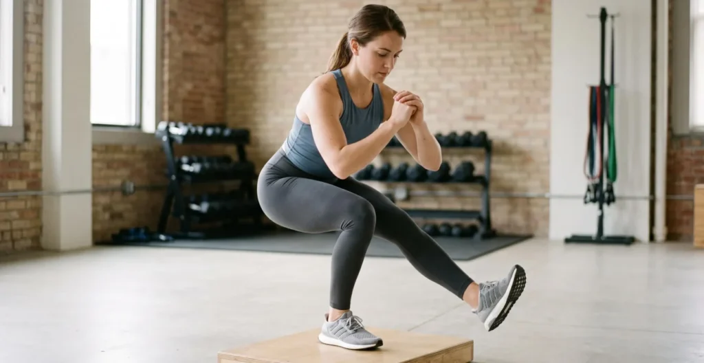
Film yourself performing a single-leg squat from a frontal view using a smartphone positioned at knee height. Stand on one leg, squat to approximately 60 degrees of knee flexion, then return to standing. Observe: Does your knee move inward past your foot’s midline? Does your pelvis drop significantly on the non-stance side? Does your trunk lean excessively? These compensations suggest hip-knee control deficits requiring targeted intervention.
For a drop jump assessment, step off a 30-centimeter box and immediately jump vertically upon landing. Film from the front and side. Evaluate landing symmetry (equal weight distribution between legs), knee flexion depth (deeper flexion indicates better shock absorption), and frontal plane knee position (maintained alignment versus valgus collapse). Failure patterns—asymmetry greater than noticeable visual difference, minimal knee bend, or clear inward knee motion—warrant consultation with a physical therapist or sports medicine professional for detailed evaluation.
Evidence-Based Neuromuscular Training Protocols
Three major prevention programs have accumulated the strongest research validation, demonstrating measurable injury reduction across multiple large-scale implementation studies. These protocols share common active ingredients—neuromuscular training combining plyometrics, strength work, balance challenges, and technique feedback—delivered in structured progressions rather than random exercise selection.
The FIFA 11+ program, originally developed for soccer but adaptable across sports, consists of a 20-minute structured warmup performed minimally twice weekly. According to National Athletic Trainers’ Association position statements, multicomponent neuromuscular training programs reduce ACL injury rates in both male and female athletes when they incorporate at least three components—strength, plyometrics, balance, agility, or flexibility—with continuous technical feedback. The FIFA 11+ integrates running patterns with directional changes (phase one), core and lower extremity strengthening combined with plyometric and balance exercises (phase two), and advanced running drills (phase three). Meta-analyses examining implementation across thousands of athletes document overall lower extremity injury reductions ranging from 30 to 50 percent when compliance reaches twice-weekly minimum frequency sustained throughout competitive seasons.
The Prevent injury and Enhance Performance (PEP) Program emerged from research in female soccer and basketball populations, emphasizing landing technique modification alongside strengthening. This 15-20 minute protocol similarly combines dynamic warmup, stretching, strengthening exercises, plyometrics, and agility drills. Validation studies demonstrate particular effectiveness in reducing noncontact and indirect-contact knee injuries, with some research showing even greater effect sizes in female populations compared to males.
Sportsmetrics represents a more intensive six-week preseason intervention (three sessions weekly) focused explicitly on ACL injury prevention through jump training, technique instruction, and strength development. Original research in female high school athletes demonstrated substantial ACL injury reduction compared to control groups, though the time-concentrated format requires dedicated training blocks rather than in-season maintenance integration.
| Program Feature | FIFA 11+ | PEP Program | Sportsmetrics |
|---|---|---|---|
| Session Duration | 20 minutes | 15-20 minutes | 60-90 minutes |
| Minimum Frequency | 2-3x per week, season-long | 2-3x per week, season-long | 3x per week, 6-week block |
| Primary Components | Running, strength, plyometrics, balance | Warmup, stretching, strength, plyometrics, agility | Jump training, strength, technique instruction |
| Implementation Timing | Pre-training warmup replacement | Pre-training warmup replacement | Dedicated preseason training phase |
| Target Population | Soccer (adaptable to multiple sports) | Soccer and basketball (female emphasis) | Female athletes, cutting/jumping sports |
| Evidence Level | Multiple meta-analyses, Grade 1A | Randomized trials, systematic reviews | Prospective cohort studies |
The convergence across these programs reveals essential principles: plyometric exercises must progress systematically from double-leg to single-leg variations and from sagittal plane (forward-backward) to frontal and transverse plane movements. Eccentric hamstring strengthening (Nordic hamstring curls, single-leg Romanian deadlifts) addresses the hamstring-to-quadriceps strength imbalance that contributes to ACL loading. Balance training progresses from stable to unstable surfaces while adding perturbations and cognitive tasks. Critically, technique feedback during execution—whether from coaches, trainers, or video review—separates effective implementation from merely completing movement repetitions.
For athletes concerned that prevention time compromises performance preparation, research indicates these protocols enhance rather than detract from athletic qualities. Improved neuromuscular coordination, reactive strength, and movement efficiency translate to performance gains in agility tests, jump height, and sprint acceleration alongside injury protection.
How does movement quality impact long-term knee resilience?
A trail runner maintaining 170-180 steps per minute generates approximately 1,000 ground contacts per kilometer. Across Marcus’s 100-kilometer training week, this totals 100,000 loading cycles that his knee structures must withstand. Even minor biomechanical deviations—several degrees of excessive valgus, five percent left-right asymmetry in force distribution, or slightly inadequate shock absorption—become magnified through this repetition volume into cumulative stress patterns that accelerate tissue adaptation demands.
This concept of cumulative biomechanical loading explains why technique optimization extends beyond acute injury prevention into long-term joint health preservation. Cartilage and ligaments respond to mechanical stress through remodeling processes, but chronic mal-distribution of forces—such as concentrated pressure on the medial knee compartment from persistent valgus positioning—exceeds the tissue’s adaptive capacity. Over seasons and years, this differential loading pattern contributes to premature cartilage degeneration, chronic ligament microtrauma, and early-onset osteoarthritic changes even without experiencing a single traumatic event.
Research examining former athletes decades after competition careers demonstrates that movement asymmetries and technical deficiencies correlate with earlier onset of degenerative knee conditions. The knee essentially ages faster when subjected to repetitive mechanical disadvantage. For endurance athletes planning multi-decade participation, investing in movement quality assessment and correction represents durability insurance as valuable as any training adaptation.
Neuromuscular fatigue amplifies biomechanical risk by degrading movement quality precisely when training stress accumulates. Studies measuring landing mechanics and cutting technique before versus after fatiguing exercise protocols consistently show deterioration—increased valgus angles, reduced knee flexion at contact, delayed muscle activation timing, and impaired postural control. This creates a paradox: the training volume necessary for performance adaptation simultaneously generates the fatigue state that elevates injury vulnerability.
Practical implications include structuring training sessions to avoid high-risk movements (explosive plyometrics, aggressive cutting drills, technical descents) during maximal fatigue states. Placing skill and technique work early in sessions, incorporating adequate recovery between high-intensity efforts, and monitoring for fatigue-related form breakdown allows athletes to accumulate necessary training loads while managing the injury risk window that fatigue creates.
Movement quality optimization also delivers performance dividends beyond injury prevention. Improved landing mechanics reduce energy dissipation, more efficient force application during push-off enhances propulsion economy, and reduced braking forces in cutting maneuvers preserve momentum. Elite athletes naturally converge on mechanically efficient patterns because inefficiency imposes metabolic costs that compromise competitive performance—the same patterns that protect joint structures.
Recovery Architecture: Sleep and Tissue Adaptation
While biomechanical optimization and training protocols receive primary focus in injury prevention discussions, the third pillar—tissue recovery capacity—determines whether accumulated training stress generates positive adaptation or progressive breakdown. Sleep represents the most critical recovery modality, during which connective tissue repair, inflammation resolution, and structural protein synthesis occur at peak rates.
During deep sleep stages and REM cycles, the body releases growth hormone and other anabolic factors that stimulate collagen synthesis in tendons and ligaments, cartilage matrix production, and muscle tissue repair. The microtrauma generated during training—normal and necessary for adaptation—requires these nocturnal repair processes to restore and strengthen tissue structures. Chronic sleep restriction interrupts this recovery cycle, leaving tissues in a partially recovered state before the next training session imposes additional stress.
Epidemiological research quantifies this relationship: a review published in Current Sports Medicine Reports indicates that athletes sleeping seven hours or less per night for periods of at least 14 days face 1.7 times greater risk of musculoskeletal injury compared to those obtaining eight or more hours. This elevated risk reflects multiple mechanisms: incomplete tissue repair, increased systemic inflammation, neuromuscular control degradation (affecting the biomechanical patterns discussed earlier), and compromised decision-making that may influence training intensity regulation.
The dose-response relationship suggests that athletes engaged in high-volume training require sleep durations at the upper end of general recommendations—eight to nine hours nightly rather than the seven to nine hours suggested for general populations. During periods of intensified training loads, such as Marcus’s preparation for UTMB qualification, sleep needs may increase further to support elevated tissue turnover demands.
Practical sleep optimization strategies for athletes include maintaining consistent sleep-wake schedules even on non-training days, creating cool bedroom environments (approximately 65-68°F or 18-20°C supports optimal sleep architecture), minimizing blue light exposure in the two hours preceding sleep, and timing intensive evening training sessions to conclude at least three to four hours before intended sleep onset. High-intensity exercise performed too close to bedtime can delay sleep onset through sustained elevation of core temperature and sympathetic nervous system activation.
Wearable sleep tracking devices (Whoop, Oura Ring, Garmin fitness watches) provide objective monitoring of sleep duration and quality metrics, allowing correlation with training load and subjective recovery perception. While these devices have measurement limitations compared to laboratory polysomnography, they establish individual baseline patterns and identify concerning trends—progressive sleep debt accumulation, unexplained sleep fragmentation, or inadequate deep sleep percentages that warrant intervention before injury risk manifests. For detailed guidance on optimizing sleep architecture for physical recovery, athletes can reference comprehensive protocols on deep sleep for physical recovery.
Beyond sleep, recovery architecture encompasses strategic rest day placement, periodization of high-load training weeks, nutrition timing to support tissue repair (particularly protein intake and anti-inflammatory nutrient patterns), and active recovery modalities that promote circulation without imposing additional mechanical stress. The integration of recovery planning with biomechanical and training protocol interventions creates the systemic prevention framework that research demonstrates achieves measurable injury risk reduction.
Medical Evaluation: When Self-Management Isn’t Enough
The prevention strategies and self-assessment protocols outlined in this article provide general educational information for athletes seeking to reduce injury risk through evidence-based training modifications. This content does not replace individualized evaluation by qualified healthcare professionals. Athletes experiencing persistent pain, functional limitations, or uncertain symptoms require clinical examination to exclude structural pathology and obtain personalized treatment planning. Self-guided prevention programs cannot diagnose existing injuries or determine appropriateness of continued training with symptomatic conditions.
When should athletes seek specialist evaluation?
Distinguishing normal training discomfort from symptoms requiring medical evaluation represents one of the most challenging decisions athletes face. The uncertainty Marcus experiences regarding his three-month anterior knee pain—wondering whether it signals adaptation versus early pathology—reflects this common dilemma. Several objective criteria help structure this decision.
Red flag symptoms warrant prompt evaluation regardless of duration. These include:
- Frank joint instability (the knee giving way or feeling unstable during normal activities)
- Significant swelling persisting beyond 48-72 hours
- Mechanical symptoms such as locking or catching that prevent complete knee extension or flexion
- Severe night pain disrupting sleep
- Traumatic injury accompanied by an audible pop or crack
- Inability to bear weight or walk normally
Any of these presentations suggests potential structural damage—ligament rupture, meniscal tear, acute cartilage injury, or fracture—requiring clinical examination and likely imaging studies.
Beyond acute red flags, temporal patterns and pain characteristics guide consultation timing. Delayed-onset muscle soreness (DOMS) typically peaks 24-48 hours post-exercise, affects muscles bilaterally and symmetrically, diminishes progressively over three to five days, and often improves with light activity or warm-up before worsening again post-exercise. This pattern represents normal physiological adaptation and does not require intervention beyond training load management.
Conversely, pain localized precisely to joint lines, tendons, or bony structures (rather than diffuse muscle distribution), persisting or worsening with continued activity rather than improving with warm-up, demonstrating clear left-right asymmetry, and failing to resolve within seven to ten days despite training modification suggests tissue irritation or injury requiring assessment. Pain that alters running or sport-specific technique—causing compensatory movement patterns—warrants earlier evaluation because continued training with compromised mechanics risks additional injury.
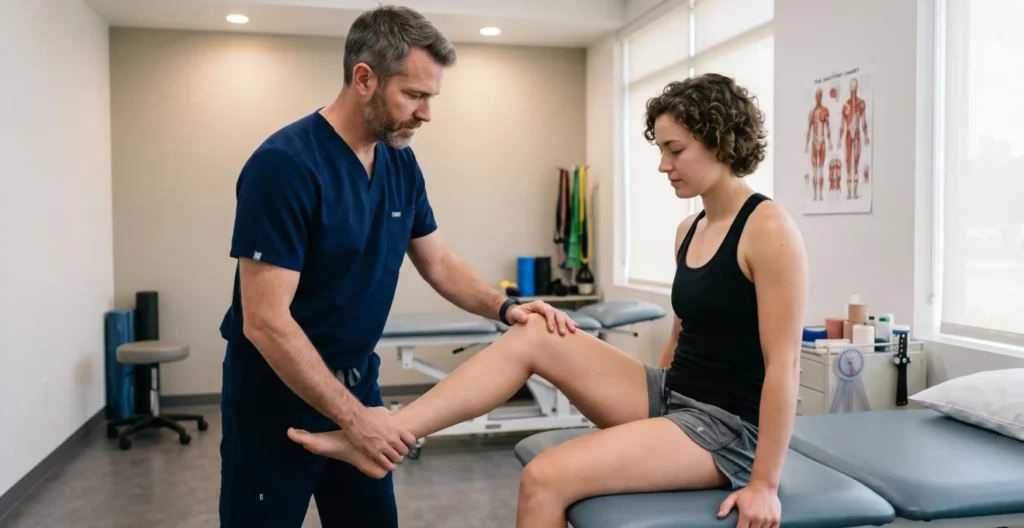
Marcus’s specific presentation—anterior knee pain recurring after long runs for three months, resolving with rest but returning with high training loads—exceeds the temporal threshold for self-management. This pattern suggests either inadequate recovery between stress applications, progressive tissue irritation (such as patellar tendinopathy or patellofemoral pain syndrome), or biomechanical loading issues requiring modification. Consultation with a physical therapist or sports medicine physician allows functional assessment, identification of contributing biomechanical factors, and structured progression planning that addresses the underlying mechanism rather than merely reducing symptoms through rest.
Within the United States healthcare system, athletes typically access specialist care through several pathways. Sports medicine physicians provide comprehensive diagnostic evaluation, can order imaging studies (MRI, ultrasound, X-ray) when clinical examination indicates structural pathology concerns, and coordinate overall treatment planning. Physical therapists offer detailed movement analysis, biomechanical screening, and conservative treatment through exercise prescription and manual therapy—many states now allow direct access without physician referral, though insurance coverage may require primary care authorization. Athletic trainers, when accessible through educational institutions or sports organizations, provide initial screening and guidance regarding appropriate referral timing.
The decision to obtain advanced imaging deserves specific mention. MRI and ultrasound are not first-line screening tools for all knee pain; their use follows clinical examination by qualified providers who determine whether imaging findings would alter treatment decisions. Research demonstrates that asymptomatic individuals frequently show meniscal tears, cartilage irregularities, and ligament signal changes on MRI without corresponding pain or functional limitation—findings that can create unnecessary anxiety and lead to inappropriate treatment if not correlated with clinical presentation. Imaging serves to confirm or exclude specific diagnostic hypotheses generated through history and physical examination, not as a substitute for clinical assessment.
Should I see my primary care doctor first or go directly to a sports medicine specialist?
Insurance networks often require primary care physician (PCP) referral for specialist coverage, making PCP consultation the practical first step for many athletes. However, some insurance plans and health systems allow direct access to sports medicine physicians or physical therapists. Check your specific coverage terms. When symptoms are clearly sports-related without systemic illness features (fever, unexplained weight loss, night sweats), direct specialist consultation provides more targeted assessment if access allows.
Can I keep training with mild knee pain?
Mild discomfort that does not alter your movement technique, resolves completely within 24 hours of training sessions, and does not progressively worsen over weeks may represent acceptable training stress. However, pain that forces compensatory movement patterns, persists into rest days, or shows cumulative worsening trend across training cycles requires load modification regardless of severity. Continuing to train through pain that alters technique risks secondary injuries from compensatory mechanics and may progress underlying tissue irritation into more significant pathology.
Do I need an MRI for knee pain?
MRI is not routinely necessary for all knee pain presentations. Many conditions—patellofemoral pain syndrome, IT band syndrome, patellar tendinopathy—are diagnosed clinically through history and physical examination without imaging. Sports medicine physicians or physical therapists order MRI when clinical findings suggest structural pathology (ligament rupture, meniscal tear, stress fracture, cartilage defect) where confirming or excluding these diagnoses would alter treatment planning. Imaging findings must be interpreted within clinical context, as asymptomatic abnormalities are common and do not necessarily explain symptoms.
How long should I wait before seeing someone about knee pain?
Acute red flag symptoms (instability, significant swelling, mechanical locking, inability to bear weight, traumatic injury with pop/crack) warrant evaluation within days regardless of pain severity. For gradual-onset pain without red flags, monitor response to training load reduction over seven to ten days. Pain persisting beyond this timeframe despite appropriate load management, pain demonstrating progressive worsening trend, or symptoms affecting normal daily activities beyond sport justify consultation. Athletes with important competition timelines (such as qualifying events) benefit from earlier evaluation to allow maximum intervention time before critical performance dates.
For athletes seeking to optimize their physiological resilience alongside structural prevention strategies, comprehensive approaches addressing respiratory efficiency can complement neuromuscular training. Resources such as the guide to nasal breathing for athletes explore how breathing mechanics influence systemic recovery capacity and training adaptation.
Building Your Integrated Prevention Framework
Effective knee injury prevention transcends isolated exercise lists to integrate three interdependent domains: biomechanical optimization identifying and correcting movement deficits through screening and technique feedback; structured neuromuscular training implementing validated protocols (FIFA 11+, PEP, Sportsmetrics) with documented 30-50 percent injury risk reduction when sustained at twice-weekly minimum frequency; and recovery architecture prioritizing adequate sleep (eight to nine hours for high-volume athletes) to support tissue repair and neuromuscular function.
The practical application begins with honest self-assessment: filming single-leg squat and landing mechanics to identify valgus, asymmetry, or landing rigidity; evaluating current sleep patterns against evidence-based duration targets; and determining whether existing training includes systematic neuromuscular work beyond sport-specific practice. Athletes identifying deficits in any domain gain clear intervention priorities rather than diffusing effort across unfocused activities.
For athletes like Marcus managing high training volumes with performance timelines, prevention integration addresses the legitimate concern that additional work may compromise specific preparation. Research demonstrates that replacing generic warm-up routines with structured neuromuscular protocols rather than adding separate sessions preserves training time while delivering protection. Movement quality work positioned early in sessions when neuromuscular control remains sharp, combined with technique emphasis during easy training days, embeds prevention within existing training architecture.
The distinction between preventable and random injury deserves final clarification. While no protocol eliminates injury risk entirely—contact injuries and unpredictable traumatic events occur regardless of preparation—the evidence unequivocally demonstrates that neuromuscular training, biomechanical optimization, and recovery adequacy substantially reduce noncontact and indirect-contact injury rates. For athletes committed to multi-year or decade-long participation, these interventions represent the difference between careers ended prematurely by preventable injuries versus sustainable progression toward long-term performance and participation goals.